
DEVELOPMENT OF SURGICAL INSTRUMENTS
Smart-Instruments Series
We developed three new surgical instruments that form as part of our Smart Instruments Series. We want to make ophthalmic surgeries smarter.
Smart-Spec Eyelid Retractor
Smart-Drape Eye Drape
Smart-Ring Pupil Dilator
Current Issues With Conventional Eyelid Retractors
Placing a Conventional Eyelid Retractor Onto The Eyelids

- With a conventional eyelid retractor, the spring and the slide structure are parallel to the arm. The hook sections open and close parallel to the eyelids.
- Therefore, even when the eyelid retractor is closed, the distance between the left and right hook section is wide. You have to tilt the device or open the eyelid with the left hand to set it on the eyelid. It is particularly difficult to set onto deep-set eyes.
Diagram of the Eyelid During the Use of a Conventional Eyelid Retractor

- When the conventional eyelid retractor is opened widely, the eyelids are lifted strongly and pain may be induced. This can also cause damage to the eyelid tissue especially damage to aponeurosis and may cause ptosis (droopy eyelid) after surgery.
- Aditionally, in the case of patients with deep-set eyes, operating the instrument proves to be difficult and water accumulation easily occurs. When the eyelids are lifted on deep-set eyes, the area becomes deeper, thus, causing instrument manipulation to be more difficult and water accumulation occurs more easily.
Solution by Mirai Eye’s Eyelid Retractor
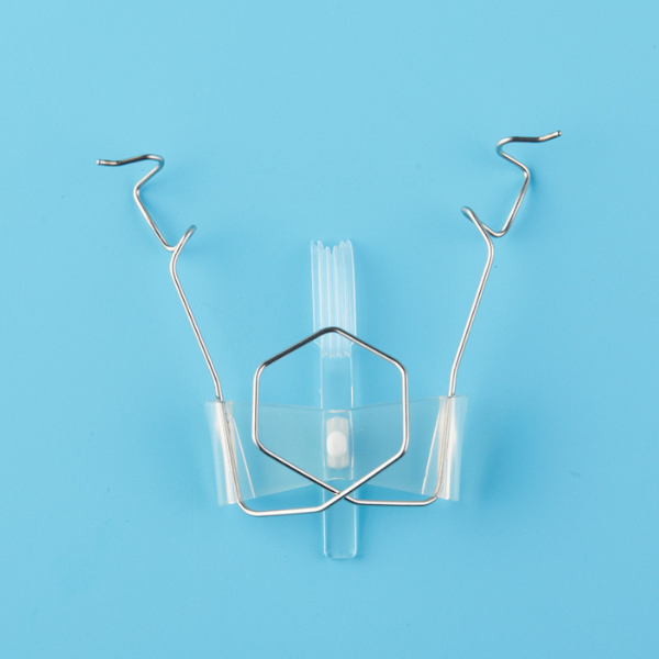
The Feature of MIRAI EYE Smart-Spec

- The Smart-Spec is an eyelid retractor which is characterized by a spring perpendicular to its arm. When its arms are closed, they are folded and the distance between the upper and lower eyelids' hooks becomes narrowed.
- Additionally, their tips also turn downward. When opened, the eyelids’ hooks open diagonally downward along the anatomy of the eyelids and eyeballs.
Placing the Smart-Spec Onto the Eyelids

- The Smart-Spec can be easily placed on most eyelids with one hand. Furthermore, since it has a structure that can hardly lift the eyelids, the eyelid tissue is less likely to be damaged.
- Simply pinch the device on the silicon section, insert the hook between the upper and lower eyelids, and then release the device.

- The Smart-Spec with a drain sheet, has a drain sheet attached to the Smart-Spec. Even with this attachment, this eyelid retractor can be easily and quickly set on the eyelids just as efficiently as the Smart-Spec with no attachment to it.
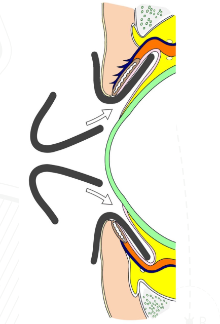
Diagram of the Eyelid During the use of a MIRAI EYE Smart-Spec
- The Insertion and removal directions of the Smart-Spec are along the curve of the conjunctival sac and eyeball. Furthermore, the hook parts have 3D curved structures along with the shape of lids and eyeball. These are the reasons that the Smart-Spec can open the lids gently without strongly pressing the lid tissues and eyeballs.
Evaluation of The Drainage Effect Using Experimental Equipment

- The drain sheet is specially grooved with two layers. This allows the drain sheet to exhibit a strong capillary phenomenon and siphon effect that strongly sucks up water and discharges it. In our experiments, we found that it surpassed the drainage capacity of conventional drainage devices.
- Since water is discharged physically, it is not necessary to connect to a suction pump like an eyelid retractor with a suction hole. This allows the conjunctiva, which is loosened by ageing, not to be sucked and therefore, the drainage effect does not decrease.
- The sheet is flat and thin and does not interfere with instrumentation, and its large width also serves as a drape to cover the exposed skin and eyelashes on the ear side.
Current Issues with Conventional Eye Drapes
- Global ageing and advances in medical technology have led to a dramatic increase in cataract surgery and intravitreal drug injections. Approximately 14 million people undergo these surgeries in Japan and the USA alone. During eye surgeries, the first thing to keep in mind is safe surgery without complications. To ensure this, accurate draping and effective drainage are extremely important.
- Accurate draping reduces the risk of developing postoperative endophthalmitis by preventing the eyelashes and skin from being exposed to the surgical field and preventing bacteria that are present on them from entering the eyeball through surgical instruments.
- With the increase in drug-resistant bacteria such as MRSA, prevention of postoperative bacterial endophthalmitis — which may lead to blindness — is a major issue for ophthalmic personnel to overcome.
- Accurate drainage reduces the risk of intraoperative complications by improving the visibility of the surgical field. It also reduces the risk for endophthalmitis to occur by discharging the bacteria which may exist in accumulated water to the outside of the eye.
Conventional Eye Drapes and Competing Products
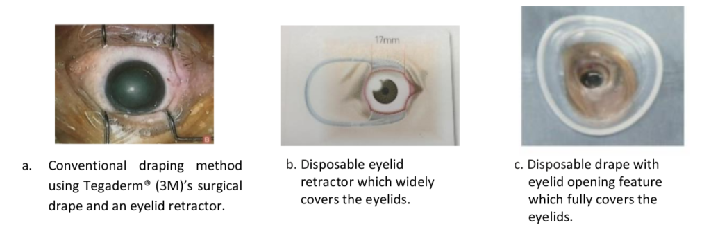
- Conventional draping methods such as Tegaderm® surgical drape and a disposable eyelid opener with a wide eyelid hook have been used. However, these methods cannot cover the ear and nose sides of the eyelids; this leaves them exposed. A disposable eyelid opener with a drape that covers the entire area of the eyelids was developed, but it did not gain popularity due to its ability to accumulate water easily.
Conventional eyelid retractor and drainage devices that drain fluids during surgery
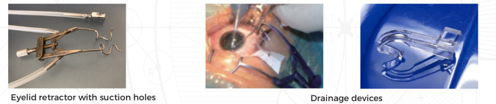
- Conventional methods to drain fluids from the surgical field include using eyelid retractors containing a suction hole that has a pump function to suck water. Others include drainage devices that are placed onto the eyelids from the ear side and drain fluids by using capillarity.
- However, there is a drawback with these — they are expensive and they cannot exert sufficient drainage functions in cases with extremely deep-set eyes or loose conjunctiva.
Solution by Mirai Eye’s Eye Drape
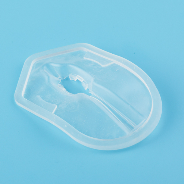
Smart- Drape

- The Smart-Drape is made up of a thin silicon film with a thickness of 0.1mm and an outer frame.
- It has a three-dimensional structure that matches the shape of the eyeball and eyelid. It has a recess at the center that is inserted under the eyelid and has a drainage structure on its ear side.
- The recess has two pockets in a vertical direction. The Smart-Spec’s hooks are inserted in a closed position to these pockets and these are fixed for use.
- The first feature of the Smart-Drape is that it can be easily and quickly set on the eyelid using the Smart-Spec.
- The second feature of the Smart-Drape is that it can reliably cover the skin and eyelashes of the eyelids all around.
- The third feature of the Smart-Drape is that it has a drainage structure on the ear side.
Placing MIRAI EYE's Smart-Drape Onto the Eyelids
- The figure below shows a series of operations on how to set the Smart-Drape on the eyelids with the use of the Smart-Spec using the cataract surgery simulator “KITARO”.

- Pinch the arms of the Smart-Spec until the distance between the left and right hooks is narrow.
- Insert them into the pockets of the Smart-Drape’s recess and join the two devices together.
- While pinched, insert this recess of the eye drape with the retractor’s hooks between the upper and lower eyelids.
- Gently release the Smart-Spec, and the pockets of recess will expand as the retractor is released. It can then be easily inserted below the eyelid.
- The recess of the eye drape that is inserted under the eyelid is covered with a silicon film all around. When the Smart-Spec is opened, the Smart-Drape stretches effortlessly according to the shape of the eyelid and eyeball.
Evaluation of Drainage Effects Using Experimental Equipment

- The figures below demonstrate the results of the Smart-Drape’s drainage function. These results were achieved by using drainage experimental equipment. When a certain amount of water is dripped from the syringe at regular intervals, the use of the Smart-Drape and Smart-Spec shows outstanding drainage in comparison to using the conventional eye drapes and eyelid retractors, which also use additional drainage devices.
- The water used during an operation is effectively drained by physical phenomena (capillary action, siphon effect, etc.). This prevents water retention and improves the visibility of the surgical field. This reduces the risk of intraoperative complications that may occur due to poor visibility. In addition to that, the bacteria that have emerged from the skin and eyelashes of the eyelids are discharged together with the water drained from the surgical field to the outside. The occurrence of postoperative bacterial endophthalmitis can be suppressed more effectively.
Current Issues With Conventional Pupil Dilators
Background and Conventional Pupil Dilators

- For cases where the pupil is not dilated with a drug (mydriatic agent), methods such as making multiple radial incisions in the pupillary margin or making a large incision in the pupil and suturing later have been performed. However, these were time-consuming and highly invasive surgical procedures.
- To solve this problem, the iris retractor was developed. For the insertion of this iris retractor, one needs to make four small incisions in the cornea and insert the iris retractor into each. Thereafter, the hook at the tip is hooked on the rim of the pupil and pulled toward the incision, and in this state, the silicone stopper is pressed against the cornea to fix it. This required considerable effort and time. In addition to this, since the hook portion is made of a thin resin wire, the pupil edge may be torn when the pupil edge is expanded by the hook, leaving the pupil deformed post-operation, which has been regarded as a problem.
- On the other hand, many ring-shaped pupil dilators have been developed to dilate the pupil more easily and in a shorter time than the above-mentioned iris retractor. These pupil dilators are naturally square or circular ring-shaped. These pupil dilators are folded and inserted into the eye with a special injector. The pupil is then expanded outward while being hooked from the inside of the pupillary edge by a groove on the outside of the ring.
Placement of conventional Pupil Dilators

- However, these pupil dilators are made of a material with high shape memory. When they are released into the eye from an injector, they are originally enlarged from a quadrangle to a circle with a diameter of about 6 to 9mm inside the eye. If you try to hook the product to the rim of the pupil, you will have to move the product widely while keeping the shape wide open. It will come in contact with the corneal endothelial cells and possibly damage them. This also strongly stretches the iris and compresses it into the iris tissue. This can result in causing damage to the iris tissue.
- Furthermore, these pupil dilators are even more difficult to insert into small pupils with a diameter of 3mm or less.
- The iris is greatly stretched and compressed. In addition to this, the product also strongly presses the inner wall of the eyeball.
Solution by Mirai Eye’s Pupil Dilator
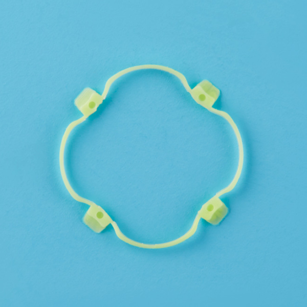
MIRAI EYE Smart-Ring
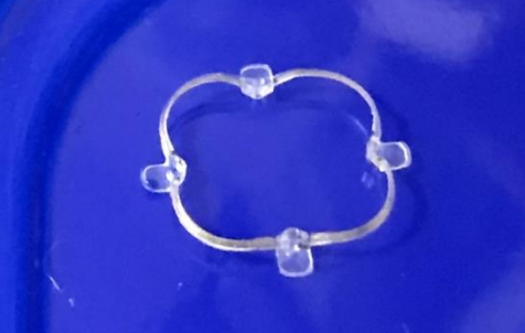
- The Smart-Ring consists of a circular ring and four iris hooks that grip the papillary margine.
- The concave sections and the convex sections of the ring are alternately arranged.
- The hook parts are located in the centre of the ring’s concave sections. These structure enables them to easily move inwards.
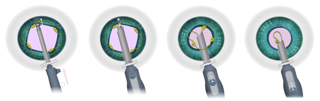
Schematic Diagram of Placing the Smart-Ring
Insertion and placement of the Smart-Ring

- Method 2: While inserting the Smart-Ring with the injector, the two-front iris-grips of the Smart-Ring will attach onto the pupil. Thereafter, attach the two-back iris-grips of the Smart-Ring using a Sinskey hook.
- Removing the insertor is done by turning it at a 90° angle clockwise so that its hook faces horizontally. This makes it easier to pull the hook out from under the ring and wound.
- The Smart-Ring is comprised of concave and convex sections. Each concave section has a iris-grip attached to it. These concave sections also easily move inwards and therefore easily grip onto the pupil without strongly stretching and compressing the iris.
Retrieval of the Smart-Ring
- Retrieval of the Smart-Ring is done by inserting the injector, and then turn it 90° clockwise so that the injector’s hook is in a horizontal position. Adjust the inserters hook so that the ring sits between the gap. In this position, turn the injector 90° anti-clockwise. Thereafter, pull the slider to suck the ring into the injector’s pipe.
- By using the methods above, we found that it is a safe, speedy and efficient way to insert and retrieve the Smart-Ring pupil dilator.